Retina Services: Diagnostic Tests
B-Scan Ultrasound
What is a B-Scan Ultrasound?
A B-scan ultrasound supplies your eye doctor with an image of the retina when he or she is unable to visualize the retina because of blood or other obstructions.Other conditions, such as a dense cataract or a cloudy cornea can block your eye doctor’s view of the retina.
The B-Scan provides an image of the back of the eye using sound emitted by a special probe such as, a submarine uses sonar sound waves to guide its way through the dark sea.
Using a clear gel probe, placed on the lids of the patient’s closed eye testing is painless lasting less than a minute.
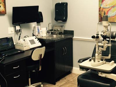
Procedure Room
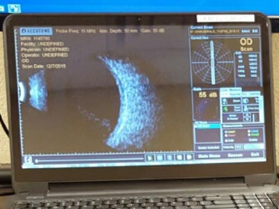
B-scan
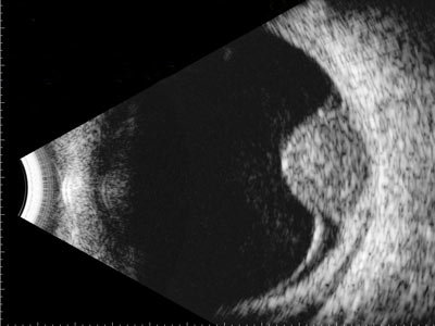
B-scan image of an iris melanoma
Why Is B-Scan Ultrasound Important in Retina Diagnostics?
B-scan ultrasonography is a critical tool in retinal diagnostics, especially when direct visualization of the retina is obstructed by conditions such as dense cataracts, vitreous hemorrhage, or corneal opacities. This non-invasive imaging technique allows ophthalmologists to assess the internal structures of the eye with precision, guiding diagnosis and treatment for a wide range of ocular conditions.
While the core function of the B-scan is to provide clear images of the posterior segment, its versatility extends far beyond basic visualization. The following section outlines additional clinical applications, patient education insights, and best practices that enhance the value of this diagnostic tool.
Q: What conditions can be evaluated using B-scan ultrasound?
A: B-scan ultrasound is commonly used to assess:
Retinal detachment (RD)
Posterior vitreous detachment (PVD)
Vitreous hemorrhage
Choroidal tumors (e.g., melanoma, hemangioma)
Optic disc drusen
Ocular trauma (e.g., lens dislocation, intraocular foreign bodies)
Posterior scleritis and uveal effusion syndrome
Q: What makes B-scan ultrasound a dynamic imaging tool?
A: B-scan provides real-time imaging, allowing the physician to observe movement patterns within the eye. This is especially helpful in distinguishing between mobile membranes (as seen in PVD) and tethered membranes (as seen in RD).
Q: Can B-scan ultrasound be used with other imaging tests?
A: Yes. B-scan is often used alongside other modalities such as Optical Coherence Tomography (OCT) or fundus photography, particularly when media opacities prevent direct visualization of the retina.
Q: What should patients expect during a B-scan ultrasound?
A: The procedure is:
Non-invasive and painless
Performed with eyes closed using a gel-covered probe
Quick, typically lasting less than a minute
Safe for all ages, including children
Requires no special preparation
Q: Is B-scan ultrasound safe, and are there any limitations?
A: Yes, it is safe and radiation-free. However:
Image quality depends on the examiner’s skill and experience
It cannot definitively distinguish between benign and malignant tumors, but it provides valuable diagnostic clues
Q: Are visual aids helpful in understanding B-scan ultrasound?
A: Absolutely. Diagrams showing probe placement, labeled B-scan images (e.g., RD, melanoma, vitreous hemorrhage), and short videos or animations can enhance patient understanding and comfort.
How does a B-Scan Ultrasound Support Advanced Retina Diagnostics?
B-scan ultrasonography is a cornerstone in ophthalmic imaging, offering detailed visualization of the eye’s internal structures when standard examination methods are limited. Its ability to penetrate media opacities—such as vitreous hemorrhage, corneal clouding, or dense cataracts—makes it indispensable in complex diagnostic scenarios.
This modality excels in identifying and characterizing posterior segment conditions, including retinal detachments, vitreous abnormalities, and intraocular tumors. In particular, B-scan imaging plays a vital role in evaluating choroidal and iris melanomas, revealing features such as dome-shaped elevation, low internal reflectivity, and posterior acoustic shadowing—hallmarks that guide differential diagnosis and treatment planning.
Unlike static imaging techniques, B-scan provides dynamic, real-time feedback, allowing clinicians to assess tissue movement and membrane behavior. This is especially useful in distinguishing between mobile vitreous strands and tethered retinal tissue.
By delivering high-resolution cross-sectional views and enabling safe, rapid assessment without radiation exposure, B-scan ultrasound enhances clinical decision-making and supports early intervention in sight-threatening conditions.


Florida
Clermont
Daytona Beach
Fernandina Beach
Fleming Island
Kissimmee
Jacksonville Riverside
Jacksonville Southside
Lady Lake
Lake City
Lake Mary
Mount Dora
Orange City
Orlando 95 Columbia St
Orlando 115 Columbia St
Palatka
Palm Coast
St. Augustine
Titusville
Wildwood
Georgia
Mondays - Fridays: 8AM to 5PM
Saturdays - Sundays: Closed
In Case of Emergency: 911
contact@floridaretinainstitute.com