
Retina Services
Retinal Vein Occlusion
What is Retinal Vein Occlusion?
A retinal vein occlusion is a blockage of one of the veins that carries blood away from the retina. The retina is delicate tissue that depends on a steady flow of blood through its arteries and veins, and when a vein becomes blocked, that circulation backs up. Patients and families often hear the condition described as an eye stroke, because like a stroke it involves a sudden interruption of blood flow.
The most common cause is one vessel pressing down on another where the two cross, narrowing the vein until it occludes. Less often, a blood clot creates the blockage.
Once a vein is obstructed, fluid leaks from the congested blood vessels into the retina, causing it to swell and thicken. That swelling is called macular edema, and it is the usual reason vision becomes blurry or is lost after an occlusion.
A retinal vein occlusion can also trigger the growth of abnormal new blood vessels. These fragile vessels may develop at the back of the eye, where they bleed, or on the iris at the front of the eye, where they interfere with fluid drainage and drive the eye pressure up. That complication is known as neovascular glaucoma, and if the pressure climbs too high it can cause pain and permanent damage to your vision.

Normal Vision:

CRVO with Macular Edema:

BRVO:
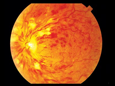
CRVO
A CRVO (Central Retinal Vein Occlusion) is a blockage of the eye's main vein (central retinal artery).
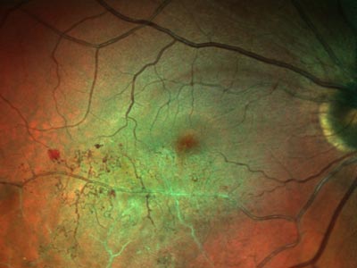
BRVO
A BRVO (Branch Retinal Vein Occlusion) is a blockage of a smaller blood vessel that branches off the main vein.
What Are the Two Types of Retinal Vein Occlusion?
Retinal vein occlusions are classified by which vein is blocked, and the distinction matters because it shapes both your symptoms and your outlook.
CRVO (Central Retinal Vein Occlusion) is a blockage of the eye's main retinal vein, where all the blood leaving the retina converges. Because the entire retina drains through this single vessel, a CRVO affects the whole retina and typically causes more significant blurring of central vision.
BRVO (Branch Retinal Vein Occlusion) is a blockage in one of the smaller veins branching off the main vein. Only the portion of the retina drained by that branch is affected, so the vision loss is more localized, often showing up in part of the peripheral field. BRVO is the more common of the two, and vision is frequently better preserved than in a CRVO.
What Causes a Retinal Vein Occlusion?
Retinal vein occlusions are usually the eye's expression of a broader vascular problem. Conditions that damage or stiffen blood vessels throughout the body make a blockage in the retina far more likely. The main risk factors are:
• High blood pressure.
• Diabetes.
• Glaucoma, or increased eye pressure.
• Blood vessel diseases and inflammatory vascular conditions.
Because these same factors affect circulation elsewhere, your eye doctor may refer you back to your primary care physician for further testing, to check whether blood vessels are blocked or at risk in other parts of your body. A retinal vein occlusion is sometimes the first visible sign of a cardiovascular problem that has gone undetected.
What Are the Symptoms of a Retinal Vein Occlusion?
The hallmark symptom is a sudden change in vision in one eye, usually without pain. Symptoms include:
• Blurriness or loss of vision, which may come on abruptly or worsen over hours to days.
• Floaters, if bleeding occurs inside the eye.
• Eye pain, which typically appears later and signals that eye pressure has risen.
If you experience any of these symptoms, it is very important to be evaluated by an ophthalmologist right away. Without treatment, a retinal vein occlusion can lead to blindness.
How is a Retinal Vein Occlusion Diagnosed?
A retinal vein occlusion is diagnosed with a dilated eye exam, in which drops widen your pupil so your doctor can examine the inside of the eye through a special lens. The pattern of retinal bleeding and swelling usually makes the diagnosis apparent.
Two imaging tests then define the extent of the damage and guide treatment:
Fluorescein angiography maps blood flow through the retina. A yellow dye called fluorescein is injected into a vein in your arm or hand, and a specialized camera photographs the dye moving through the retinal vessels. The images reveal where circulation has been cut off and whether abnormal new vessels are forming.
Optical coherence tomography (OCT) scans the retina to produce detailed cross sections, like a virtual biopsy. OCT is how your doctor measures macular edema precisely and tracks whether swelling is responding to treatment.
How is a Retinal Vein Occlusion Treated?
Treatment depends on the extent of the damage, and it has two parallel aims: reducing the swelling that is blurring your vision, and preventing the complications that threaten it longer term.
Medication injections. Anti-VEGF medications injected into the eye are the primary treatment for the macular edema caused by a vein occlusion. They reduce leakage from congested vessels, allowing the swelling to subside and vision to improve. Steroid medications are another option for reducing edema. Injections are typically repeated over time, and your retina specialist will adjust the schedule based on how your eye responds.
Laser treatment. Laser may be used to treat persistent swelling in some cases, and it plays an important role when abnormal new blood vessels develop. Panretinal photocoagulation reduces the stimulus driving that growth and helps prevent neovascular glaucoma.
Managing your overall health. Controlling the conditions behind the occlusion, particularly high blood pressure and diabetes, is a genuine part of your eye treatment. It protects the affected eye from further damage and lowers the risk of an occlusion developing in your other eye.
Frequently Asked Questions About Retinal Vein Occlusion
Is a Retinal Vein Occlusion an Emergency?
Yes, it should be treated as urgent. Sudden painless vision loss in one eye needs same day or next day evaluation by an eye doctor, because early treatment of macular edema gives you the best chance of recovering vision, and because complications like neovascular glaucoma are far easier to prevent than to reverse. Sudden vision loss can also indicate a retinal artery occlusion, a related but more emergent condition, which is another reason not to wait and see.
Will My Vision Come Back After a Retinal Vein Occlusion?
Often, at least partially. Many patients regain useful vision once macular edema is treated, and outcomes tend to be better with a BRVO than a CRVO, and better when treatment begins early. How much vision returns depends on how much of the retina lost its blood supply and on whether the central macula was affected. Some people recover most of their sight, while others are left with lasting blurring. Your retina specialist can give you a more specific picture after reviewing your imaging.
Can a Retinal Vein Occlusion Happen in the Other Eye?
It can. Having one occlusion indicates that your blood vessels are vulnerable, which raises the risk in the fellow eye compared with someone who has never had one. This is precisely why controlling blood pressure, blood sugar, and other vascular risk factors matters so much after a diagnosis, and why your retina specialist will examine both eyes at every visit.
How is a Retinal Vein Occlusion Different From an Eye Stroke or a Retinal Artery Occlusion?
The phrase eye stroke is used loosely for both conditions, which causes confusion. A vein occlusion blocks blood leaving the retina, so circulation backs up, and vision loss usually develops over hours to days. An artery occlusion blocks blood arriving at the retina, starving the tissue of oxygen, and it causes profound vision loss within seconds to minutes. An artery occlusion is a true emergency closely linked to stroke risk elsewhere in the body and requires immediate evaluation. Either way, sudden vision loss warrants an urgent call.
What Lifestyle Changes Help After a Retinal Vein Occlusion?
The most valuable changes are the ones that protect your blood vessels, since a vein occlusion is fundamentally a vascular event. Working with your primary care doctor, that generally means:
• Keeping blood pressure in your target range, which is the single most important factor for most patients.
• Controlling blood sugar if you have diabetes.
• Managing cholesterol and having your circulation evaluated.
• Stopping smoking, which damages blood vessels throughout the body.
• Staying physically active and eating a diet built on vegetables, whole grains, and lean proteins.
• Taking prescribed medications consistently, including any blood pressure or blood thinning medication your doctor recommends.
None of this reverses an occlusion that has already occurred, but it meaningfully lowers your risk of another one and supports the health of the eye you are treating.
Does a Retinal Vein Occlusion Cause Blindness?
It can if left untreated, particularly when neovascular glaucoma develops and eye pressure damages the optic nerve. With prompt diagnosis and modern treatment, however, complete blindness is uncommon. Most patients who are treated retain useful vision, which is why an urgent evaluation and consistent follow up matter so much.
If you have experienced sudden blurring or loss of vision in one eye, do not wait. Please contact Florida Retina Institute or call our office right away to speak with a member of our team. With 21 locations throughout Florida and Georgia, expert retina care is always close to home.



Florida
Clermont
Daytona Beach
Fernandina Beach
Fleming Island
Kissimmee
Jacksonville Riverside
Jacksonville Southside
Lady Lake
Lake City
Lake Mary
Mount Dora
Orange City
Orlando 95 Columbia St
Orlando 115 Columbia St
Palatka
Palm Coast
St. Augustine
Titusville
Wildwood
Georgia
Mondays - Fridays: 8AM to 5PM
Saturdays - Sundays: Closed
In Case of Emergency: 911
contact@floridaretinainstitute.com