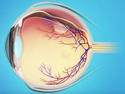
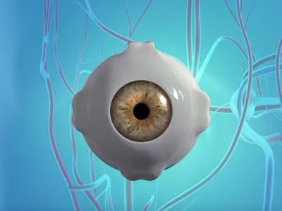
Retina Services
Vitreo-Retinal Diseases and Surgery
Excellence in the Treatment of Vitreo-Retinal Diseases and Surgery since 1979
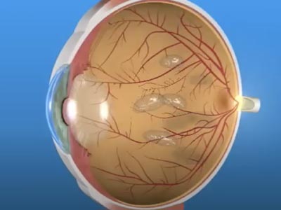
Retinal Detachment
A detached retina or retinal detachment is when the retina peels away from the back wall of the eye. The retina does not function properly when it is detached, resulting in seeing a peripheral shadow that can progress to cause significant blurring of the vision.
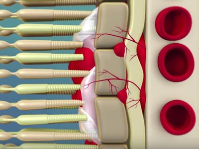
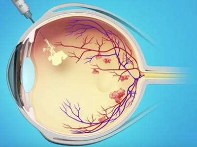
Diabetic Retinopathy
Diabetic retinopathy is a complication of diabetes caused by high blood sugar levels damaging the tiny blood vessels in the retina. Over time, these vessels can weaken, swell, and leak fluid or blood, leading to vision changes and, if untreated, potential vision loss.
Macular Degeneration
Age-Related Macular Degeneration (AMD) is a condition that damages the central part of the retina, leading to difficulty with tasks like reading, recognizing faces, and seeing fine details. It mainly affects older adults and causes gradual loss of central vision.
Vitreous and Macular Surgery
Vitreous and Macular Surgery treats conditions affecting the retina, macula, and vitreous—structures vital for sharp, central vision. These minimally invasive procedures help restore sight and address issues like Macular Hole, Macular Pucker, and Floaters and Flashes.
Macular Hole Repair
A macular hole is a small break in the macula, a hole or defect that develops in the center of your eye’s light-sensitive tissue, called the retina. With the development of a macular hole, your central vision will become blurry, wavy or distorted.
Floaters and Flashes
Vitreous Floaters and Flashes are common symptoms caused by changes in the gel-like vitreous inside the eye. Floaters appear as drifting specks or threads and result from clumps of gel casting shadows on the retina. Flashes look like brief bursts of light and occur when the vitreous pulls on the retina, often with age.
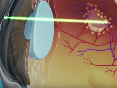
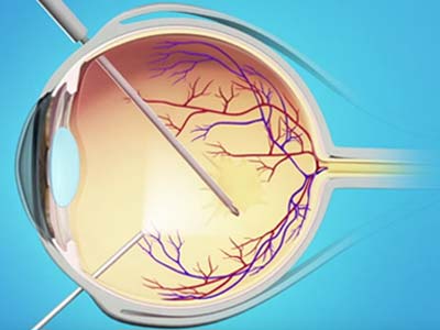
Laser Procedures
A laser is strong beam of light. An ophthalmologist uses different types of lasers to perform a variety of laser procedures. Two different kinds of lasers are used in eye surgery. Your retina specialist will primarily use the thermal laser.
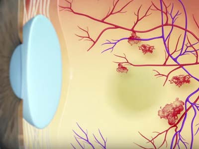
Retinal Vein Occlusion
The retina is sensitive tissue that requires blood flow from arteries and veins. A blockage or occlusion of a vein results in a retinal vein occlusion. The most common cause of a blockage is one vessel pressing down on another and causing it to narrow and occlude.

Pediatric Retina Conditions
Pediatric retina encompasses the common eye disorders of childhood, such as retinopathy of prematurity. Retinopathy of prematurity occurs when babies are born prematurely and the blood vessels of the retina inside the eye do not develop properly.
Diagnostic Tests
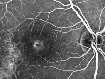
Fluorescein Angiography
Retinal Angiography is a diagnostic procedure that will enable your doctor to visualize the blood vessels and tissue of the eye. By examining these tissues more closely with the pictures your ophthalmologist can diagnose many eye diseases and keep track of changes over time.
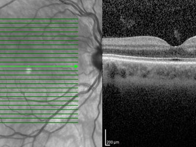
Optical Coherence Tomography
Optical Coherence Tomography (OCT) is a commonly performed diagnostic test designed to assist your doctor in identifying retinal diseases, such as age-related macular degeneration or diabetic eye diseases.
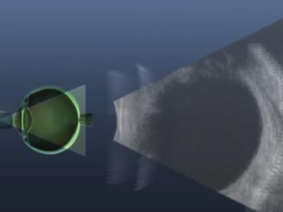
B-Scan Ultrasound
A B-scan ultrasound supplies your eye doctor with an image of the retina when he or she is unable to visualize the retina because of blood or other obstructions.


Florida
Clermont
Daytona Beach
Fernandina Beach
Fleming Island
Kissimmee
Jacksonville Riverside
Jacksonville Southside
Lady Lake
Lake City
Lake Mary
Mount Dora
Orange City
Orlando
Palatka
Palm Coast
St. Augustine
Titusville
Wildwood
Georgia
Mondays - Fridays: 8AM to 5PM
Saturdays - Sundays: Closed
In Case of Emergency: 911
contact@floridaretinainstitute.com