Retinal Diseases, Laser Procedures and Surgery
Diagnostic Tests
About Fluorescein Angiography
What is a Retinal Angiography?
Retinal Angiography is a diagnostic procedure that will enable your doctor to visualize the blood vessels and tissue of the eye. By examining these tissues more closely with the pictures your ophthalmologist can diagnose many eye diseases and keep track of changes over time.
What happens during Retinal Angiography?
Retinal Angiography is usually done in your ophthalmologist's office.
Capturing the image takes less than 30 minutes.
Here is what to expect:
The ophthalmologist’s assistant will put drops in your eyes to dilate (widen) your pupil.
A colored dye (yellow), called fluorescein is injected in a vein, usually in your arm.
It takes about 10 - 15 seconds for the dye to travel to the eye.
The yellow dye highlights the blood vessels and enables your eye doctor to look at your choroid and retina.
As the dye passes through your retina and choroid, an ophthalmic technician takes a series of preliminary photographs using a special camera, called the fundus camera.
These pictures become a roadmap to help your ophthalmologist see where to focus treatment if a disease is detected.
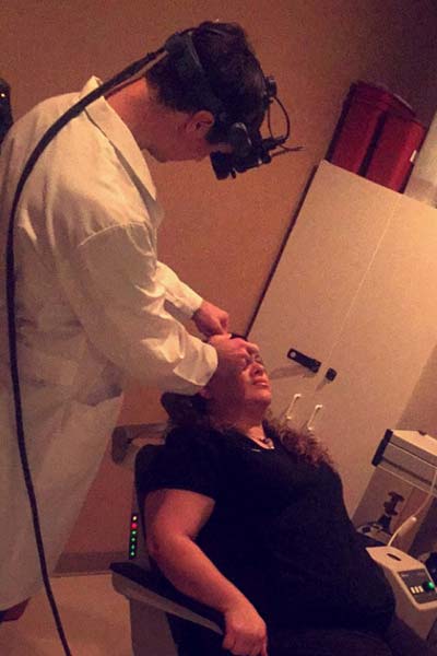
Dr. Steven Houston with a patient.
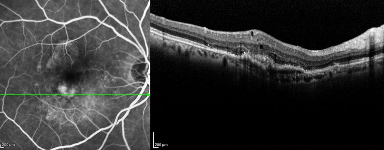
In these images, the macular hole and areas of active wet macular degeneration are highlighted by the fluorescein dye, which circulates through the blood vessels in the back of the eye.
What to expect after Retinal Angiography
If possible, please have a driver accompany you to the doctor's office.Your vision will be immediately affected by the bright lights of the camera. This will resolve but your vision will stay blurry for several hours from the eye drops used to dilate your pupils.
Also, your eyes will be very sensitive to light. Wearing sunglasses after your test will make you more comfortable.
Retinal Angiography Risks and Side Effects
Your skin may take a yellow appearance. This happens because the dye travels to all your veins in your body. Your skin will return to normal pigmentation in a few hours.Your urine may look orange or bright yellow for up to 24 hours after angiography because your kidneys must filter the dye out of your blood.
You may feel a burn on your skin if dye leaks during the injection. The sensation should subside in a few minutes.
Rarely, allergic reactions to the dye occur. People who are allergic to the fluorescein dye may get hives or itchy skin. Very rarely, a person may have breathing or other serious problems. Please, tell your doctor if you have had previous reactions with yellow dye used in angiography.
Allergic reactions can be treated by your doctor with the use of medication.
Retinal angiography is a diagnostic procedure
that involves taking detailed images of the retina and its blood circulation.
Using these pictures, the eye doctor can monitor your retina and other parts of the eye for any leakage or abnormality.
A colored dye is injected into your arm, where it travels through blood vessels to your retina. Using a special camera, pictures of the retinal vessels glow with dye.
Your eye doctor can diagnose problems using this test and the images as a roadway to guide treatment.
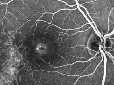 This image shows evidence of a macular hole.
It is highlighted by the fluorescein dye, circulating through the blood vessels of the retina.


Florida
Clermont
Daytona Beach
Fernandina Beach
Fleming Island
Kissimmee
Jacksonville Riverside
Jacksonville Southside
Lady Lake
Lake City
Lake Mary
Mount Dora
Orange City
Orlando 95 Columbia St
Orlando 115 Columbia St
Palatka
Palm Coast
St. Augustine
Titusville
Wildwood
Georgia
Mondays - Fridays: 8AM to 5PM
Saturdays - Sundays: Closed
In Case of Emergency: 911
contact@floridaretinainstitute.com