Retina Services: Diagnostic Tests
Optical Coherence Tomography
What is Optical Coherence Tomography or OCT?
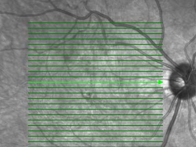
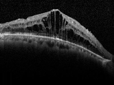
Optical Coherence Tomography (OCT) is a commonly performed diagnostic test designed to assist your doctor in identifying retinal diseases, such as age-related macular degeneration (AMD) or diabetic retinopathy (diabetic eye disease).With an Optical Coherence Tomography (OCT) we can look closely at the retina and obtain a virtual, non-invasive biopsy. Similar to a CAT scan, the OCT uses infrared light rather than x-rays to form a cross-sectional view of your retina. This detailed image of the retina allows your doctor to see the retina at a microscopic level and detect swelling in the retina.
"Tomos" means section
Non-invasive Imaging
Infrared Light
Are you Preparing for an OCT Exam?
What you should expect during an Optical Coherence Tomography (OCT) exam: Your doctor may put dilating eye drops in your eyes in order to widen your pupil and make it easier to examine the retina.
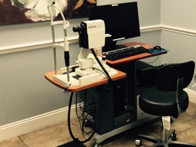
After the pupils are dilated, the patient is seated in front of the OCT machine, the head rests on a support to keep it motionless.
The patient simply looks into the lens of the device at a small, blinking target, the equipment scans the eye without touching it.
The OCT test is over in seconds. If your eyes were dilated, they may be sensitive to light for several hours after the exam.
Detailed Diagnosis
Treatment and Monitoring of your Retinal DiseaseThe Optical Coherence Tomography technology provides great components for examining the vitreo-retinal interface, as it helps to pinpoint retinal pathologies and detect retinal edemas.
The OCT uses data to monitor the progression of the disease and with it your doctor can determine if treatment is necessary.
It also enables your eye doctor to tell whether or not retinal therapies such as laser or eye injections are decreasing the fluid or edema in the retina.
Expect to get an OCT test on almost every visit to your retina doctor.
What is the Heidelberg Spectralis?
The Heidelberg Spectralis® is a high definition, spectral domain optical coherence tomographer by Heidelberg Engineering.The Spectral (or Fourier) Domain OCT (SD-OCT) uses a significantly faster, non-mechanical technology.
 The SPECTRALIS® SD-OCT simultaneously measures multiple wavelengths of reflected light across a spectrum, hence the name spectral domain. The SPECTRALIS system is 100 times faster than TD-OCT and acquires 40,000 A-scans per second.
The increased speed and number of scans translates into higher resolution and a better chance of observing disease.
Moving at the Speed of Light
The Spectralis® system is much more than just a faster next generation OCT.
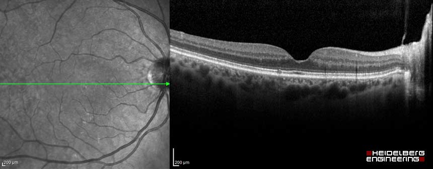
High speed image acquisition is combined with custom TruTrack™ technology to actively track the eye during imaging. Tracking eye movement with simultaneous dual-beam imaging minimizes motion artifact, enables noise reduction and allows the instrument to precisely track change over time. The result is point-to-point correlation between fundus and OCT scans, greater image detail and clarity, and a more confident assessment of small changes.